Lab Notes
✶ March 10, 2026 ✶
Viewing biofilm chunks and aquatic fungi (debateable - because I'm not an expert).
New Unknowns: The Witch and the Nematode-Trapping Fungi
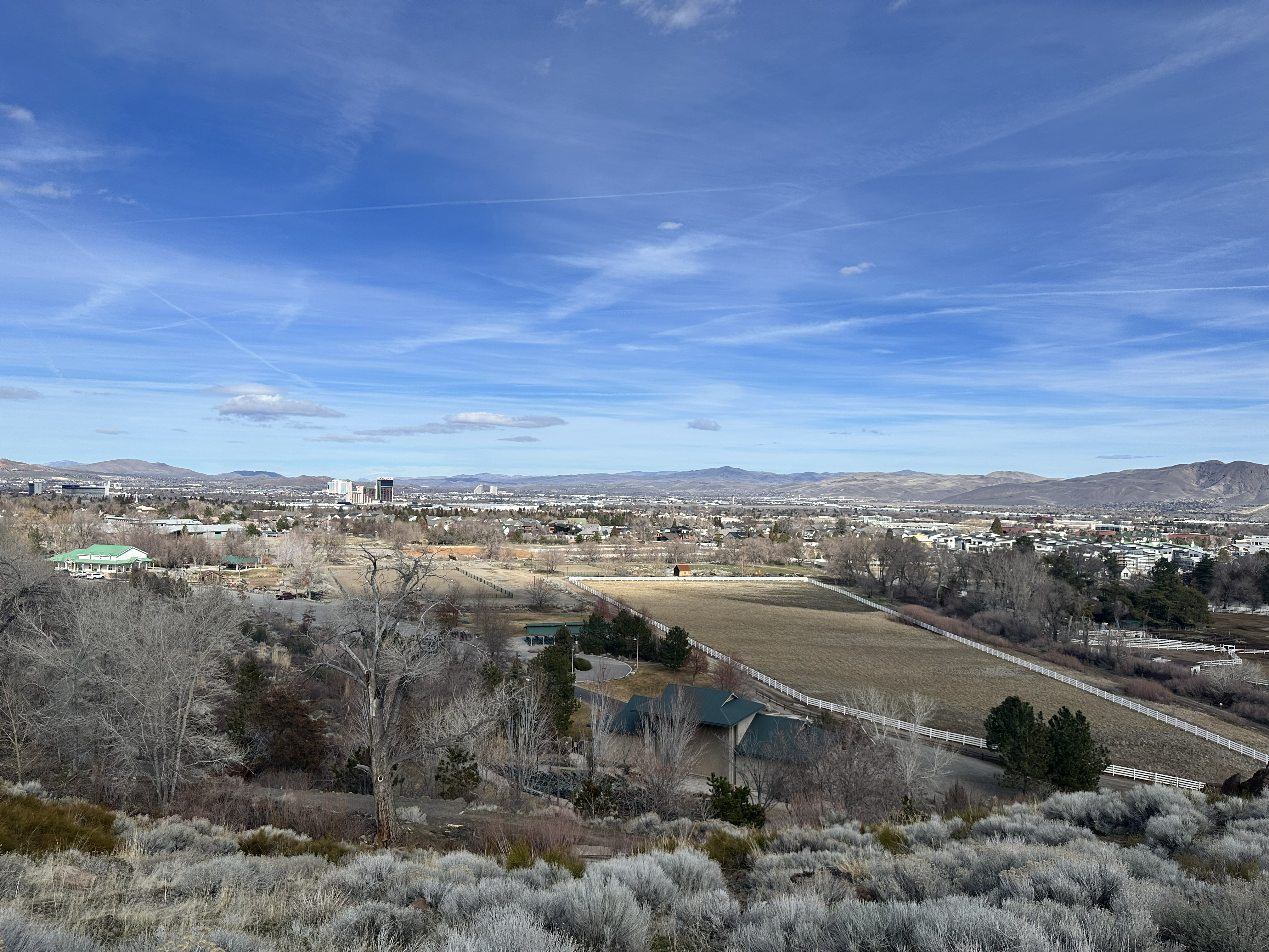
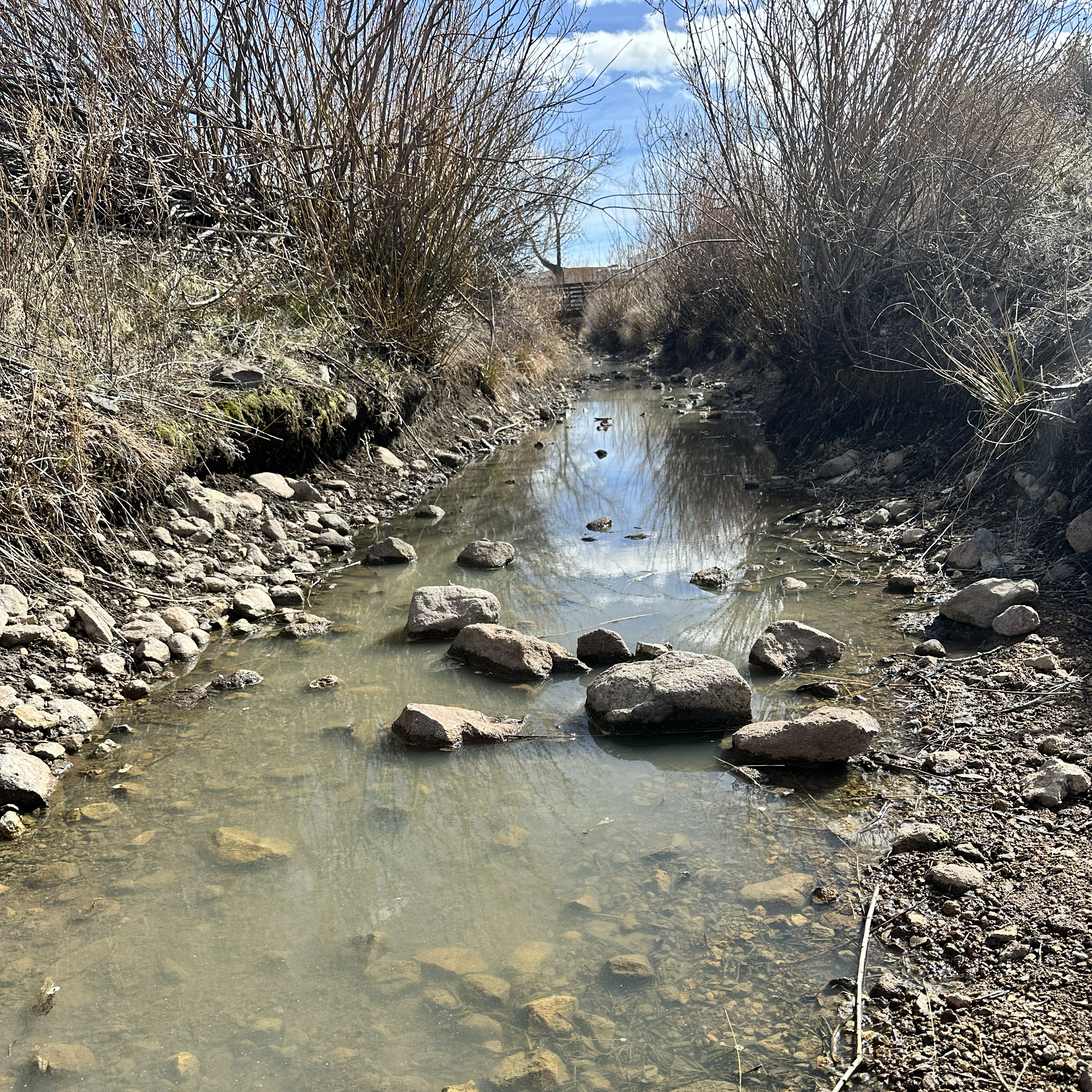
We had our fun with the running stream water last time, so now: the stagnant water. We got the sample from the same park at the same time as the Evan's Creek sample. In fact, the stagnant water came from an old agiculture waterway, Lake Ditch, that supported the park when it was still a dairy farm before WWII. It was later turned into a horse ranch and park by the owner's son, and the waterway dries up here and there throughout the year. When I collected the sample, we were nearing the end of winter, but before any real amount of snow runoff would increase its volume. Regardless, I did find a small ecosystem full of different things: bacteria, organic debris, and filamentous fragments. Now, I'm not an expert - far from, as this is simply a hobby I enjoy. So take my findings with a grain of salt.
Experiments: Swift 380T Microscope @ 400x & 1000x magnification
| Organism | Observations | Photos |
|---|---|---|
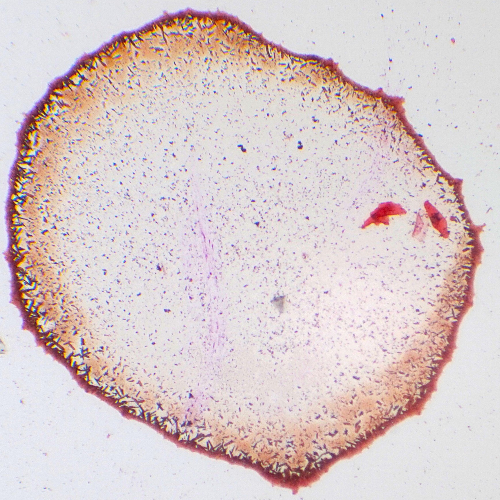
Biofilm AggregateUnknown genus/species |
Before anything, this sample was Gram stained: hence some pinkish coloring that normally wouldn't be there. This was also one of the first things I found upon inspection. I went straight to research, as I was pretty stumped on what this was. From said research, I was able to faily conclude that it's a just a chunk of biofilm, acting as a mini-city for numerous different microorganisms. You can see in the close-up image that there's many bacteria that are clinging to the edges along with filament fragments. Now, I'm not able to really tell what type of bacteria this is within the chunk, however, I feel like two good options are: Pseudomonas or Aeromonas-type bacteria. These two are good contenders due to their Gram-negative (pinkish) stain and their aquatic environments with stagant fresh water being one of them. Psuedomonas also form biofim and appear as tiny pink rods, while Aeromonas like to live in biofilms along with algae and other cyanobacteria. Unfortunately, I don't have the tools nor expertise to find out their exact genetic makeup and narrow it down from there. There is a post in the Grimoire for the Gram staining technique I used to get this result as well. I would like to experiement with negative staining next if possible. I would also like to note that I did see many other fragmented pieces floating around, like tiny shards when you break a chip or plate. Also, due to the staining, the bacteria in this sample are all dead (alcohol is used to keep the organisms adhered to the slide, but it also kills most of them). This makes it difficult to know the motility of anything in this area. |
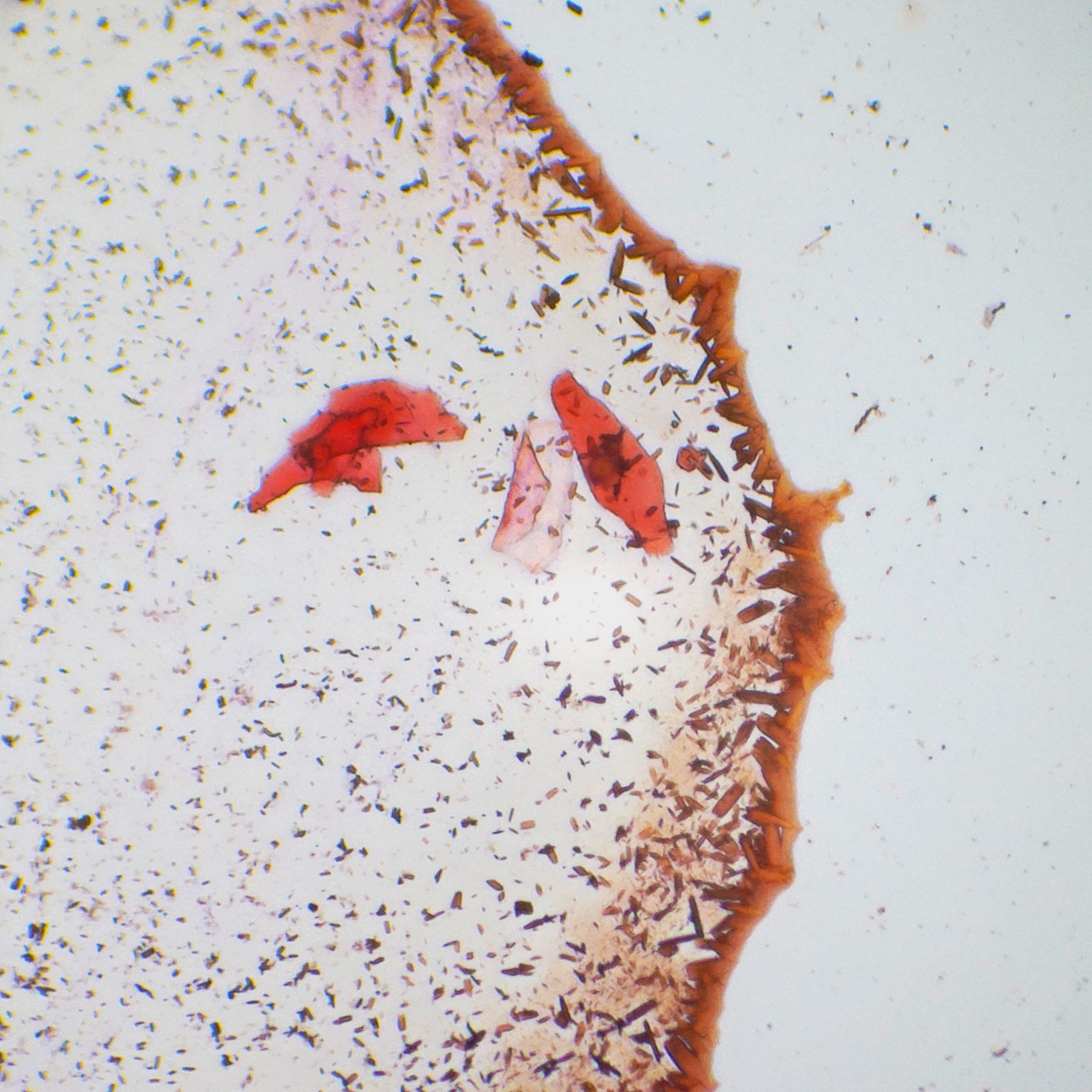 |
Nematode-trapping fungiNematophagous Fungi |
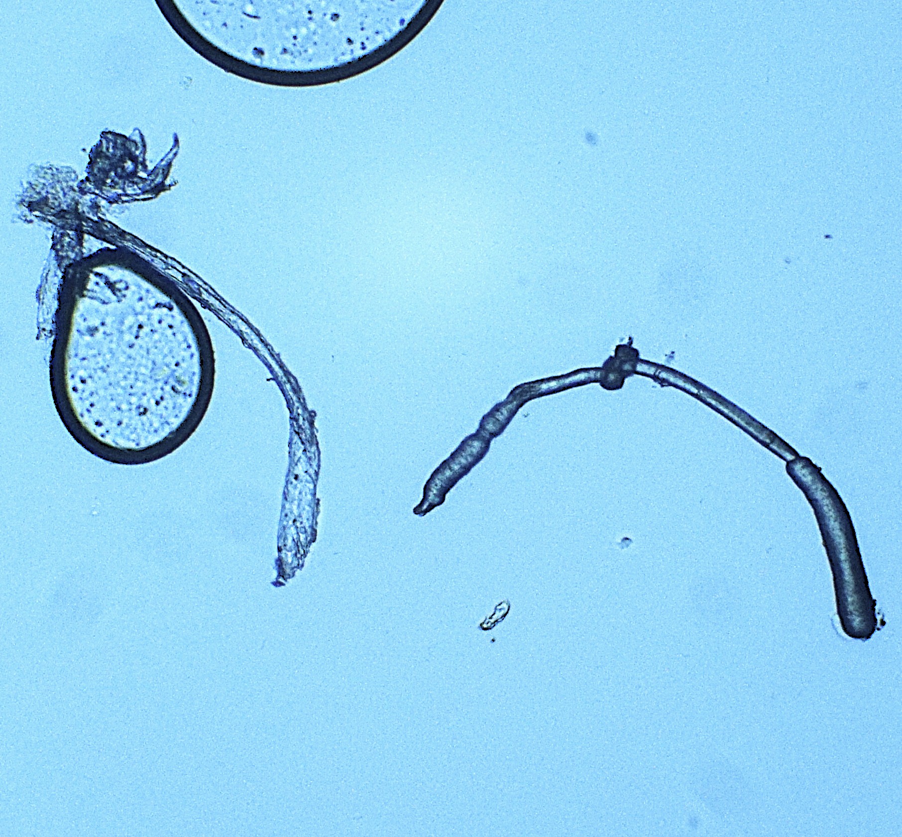
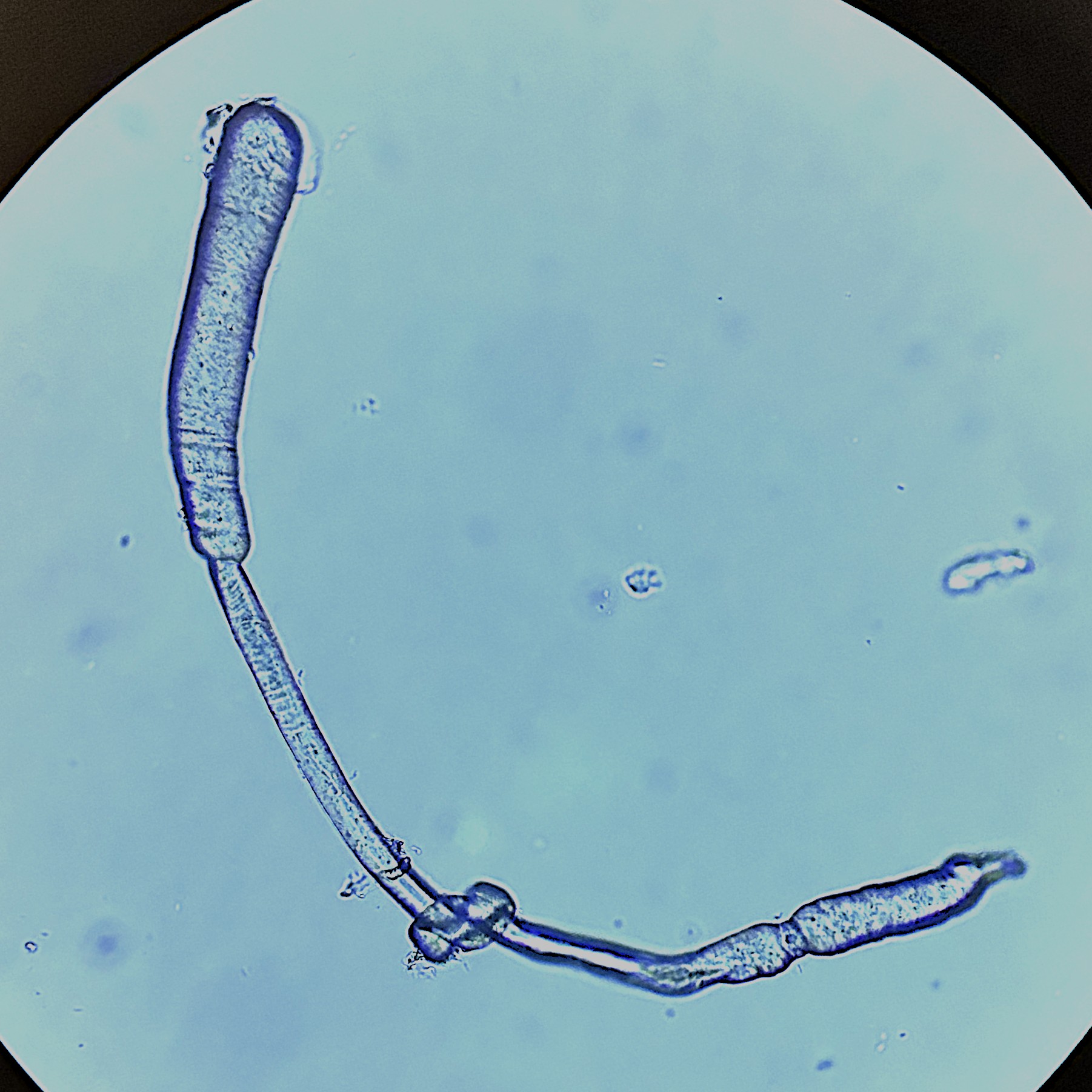
As with the biofilm chunk above, I can't say with certainty of what we're looking at. At first glance, I thought, "Cool! A worm!" But then quickly came to the conclusion that it was indeed not a worm. One, it was lacking the basic worm-like structures such as a mouth, organs, or even differentiated tissues, as well as too small. So the next best idea was that it was a fragmented piece as well, but the shape wasn't linear enough. And after quite a bit more digging the best conclusion I can find is Nematode-trapping fungi. Because it clearly has a "head" and "tail" end that are more like swollen nodes, it gave off a more fungal mood. The center part that looks clumped actually looks to have been knotted around itself (like a shoelace). It's body is also fairly transparent with no sign or internal organs. A quick reminder that this was found in stagnant freshwater in Northern Nevada, it's known that nematodes (roundworms) can make their home here. But checks-and-balances play a part everywhere, even in nature. And the nature predator of the nematode is the Nematode-trapping fungi. This fungi has hyphal filaments, long transparent tubes, that grow to produce traps for the nematodes. These act like fingers and are able to grab and constrict nematodes by knotting around them. Almost exactly as seen here. We are, however, missing the rest of the network for confirmation - as these fungi appear in vast networks almost like spiderwebs. It's still entitrely possible that I broke this piece off while obtaining the sample and preparing it on the slide. |
 |
Location of sample
Bartley Ranch Regional Park (Reno, NV)